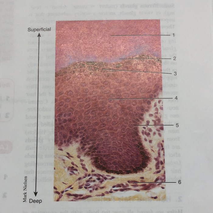
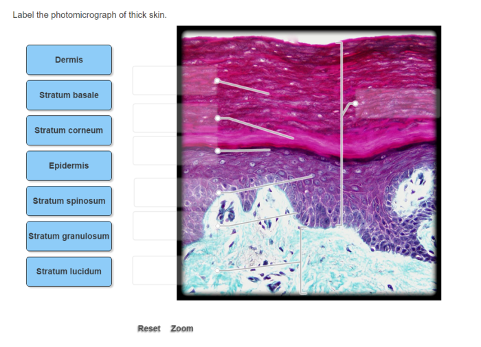
Label the photomicrograph of thick skin. – Labeling the photomicrograph of thick skin is a crucial technique in histology and pathology. It involves identifying and labeling the key structures visible in the photomicrograph, providing valuable information for diagnostic purposes, educational materials, and research studies.
This guide will provide a comprehensive overview of the labeling process, including the key structures to identify, the labeling techniques used, and the applications of labeled photomicrographs in various fields.
Labeling the Photomicrograph of Thick Skin

Thick skin is a specialized type of skin found in areas of the body subjected to friction and pressure, such as the palms of the hands and soles of the feet. It consists of three main layers: the epidermis, dermis, and hypodermis.
Labeling a photomicrograph of thick skin helps identify and visualize these layers and their constituent structures.
Key Structures
- Epidermis:The outermost layer of thick skin, consisting of multiple layers of keratinized cells that provide protection against wear and tear.
- Dermis:The middle layer of thick skin, composed of connective tissue, blood vessels, hair follicles, and sweat glands.
- Hypodermis:The innermost layer of thick skin, consisting of adipose tissue that insulates and cushions the body.
Labeling Techniques

Various labeling techniques are employed to identify specific structures in a photomicrograph of thick skin, including:
- Immunohistochemistry:Uses antibodies to bind to specific proteins within the tissue, allowing visualization of the distribution of those proteins.
- Histochemical staining:Uses dyes or stains to react with specific molecules or structures within the tissue, providing contrast for visualization.
- Digital image analysis:Employs computer software to enhance and analyze digital images of tissue sections, facilitating the identification of specific structures.
Each technique has its advantages and disadvantages, and the choice of technique depends on the specific structures of interest and the desired level of detail.
Example of a Labeled Photomicrograph
 |
|
Applications of Labeling: Label The Photomicrograph Of Thick Skin.

Labeling photomicrographs of thick skin has several applications, including:
- Histological studies:Examining the structure and organization of thick skin in different physiological and pathological conditions.
- Diagnostic purposes:Identifying specific skin diseases or abnormalities based on the presence or absence of certain structures.
- Educational materials:Illustrating the anatomy and histology of thick skin for educational purposes.
FAQ
What are the key structures visible in a photomicrograph of thick skin?
The key structures include the epidermis, dermis, and hypodermis.
What labeling techniques are commonly used for photomicrographs of thick skin?
Common labeling techniques include hematoxylin and eosin staining, immunohistochemistry, and fluorescent labeling.
What are the applications of labeled photomicrographs of thick skin?
Labeled photomicrographs are used in histological studies, diagnostic purposes, and educational materials.
